|
SUMMARY
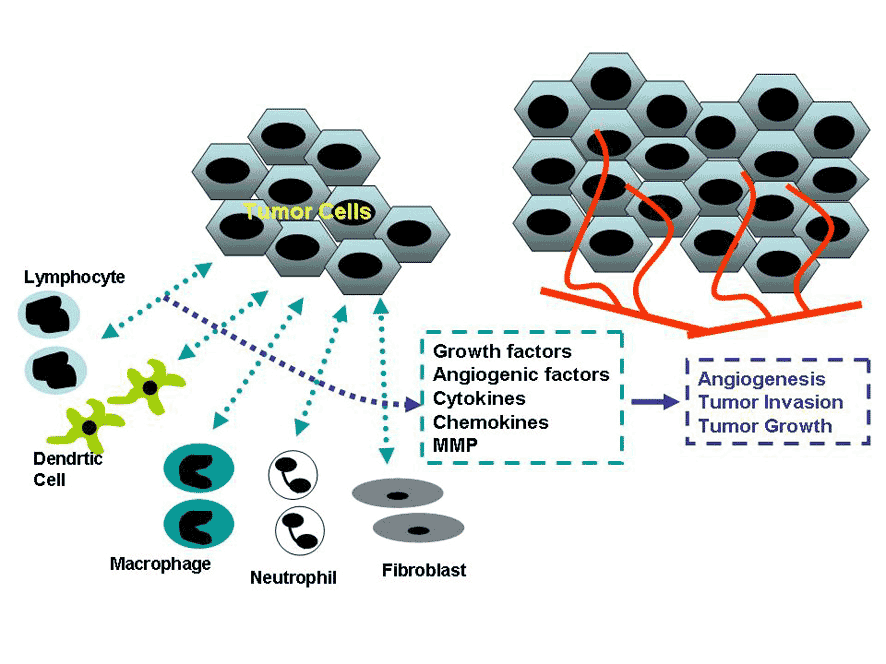
ü@Upon the tissue injurise arising from several causes including microbial infection and trauma, mammals including humans exhibit host protective responses, so-called inflammatory responses, characterized by the infiltration and activation of leukocytes, the proliferation of fibroblasts and neovascularization. Usually, these responses eventually result in the reduced tissue damages. ü@During the course of inflammatory responses, animals produced various inflammatory cytokines including chemokines, growth factors, matrix metalloproteinases (MMPs), reactive oxygen species (ROS), and angigenic factors, along with the release of several enzymes such as cyclooxygenases (COX). ü@Evidence is accumulating to indicate that chronic infection witb Helicobacter pylorii and hepatitis viruses B and C, have crucial roles in the development of
gastric cancer and hepatoma, respectively. Moreover, because these microbes
lack proto-oncogenes at all, chronic inflammation caused by these microorganisms
is presumed to be essentially involved in the carcigenesis of these cancers. ü@In most types of tumors except leukemia, tumor cells co-exist with stroma
cells such as fibroblasts and endothelial cells. Moreover, tumor tissues
attract various types of leukocytes including granulocytes, monocytes/macrophages,
lymphocytes, and dendritic cells accumulate inside the tumor lesions, because
these cells recognize tumor cells as forein cells and exhibit inflammatory
responses. Thus, tumor tissues, particularly solid tumor tissues, are formed
under the interaction between tumor cells, and stroma cells and leukocytes.
Figure Interaction between tumor cells and fibroblasts and leukocytes ü@Based mainly on in vitro studies, evidence is accumulating to indicate that stroma cells and leukocytes can produce inflammatory cytokines including chemokines, growth factors, MMP, ROS, and angiogenic factors, and release enzymes such as COX, under the interaction with tumor cells. The produced molecules can act on tumor cells themselves and induce neovascularization, an essential component for the nourishment of tumor tissues. These processes can eventually develop tumor progresssion, leading to metastasis. ü@In order to develop control and/or inhibit the progression and metastasis of tumors, it is mandatory to determine the pathophysiological roles of these molecules more in detail. For this purpose, it has become necessary to examine their roles using animal models as well as in vitro studies. ü@In order to investigate the roles of these molecules in the development
of tumor formation, our group is using several metastasis and carcinogenesis
models, together with the use of mice deficient in pro-inflammatory cytokines
and chemokines. ü@Our major achievements over the past three years can be summarized as follows. ć@ü@In normal bone marrow, basophils constitutively produce a chemokina, CCL3, which inhibit the proliferation of normal hematopoietic stem cells, particularly at bone marrow transplantation. In chronic myeloid leukemia, an increase in basophil-like leukemia cells are reported for a long time. CCL3, produced by basophil-like leukemia cells, suppressed the growth of normal hematopoieitc stem cells, thereby favoring the growth of leukemia-initiaing cells and subsequent leukemia development (Baba et al., 2013; Baba et al., 2016) . ćA Chemokines, produced in tumor sites, induces the accumulation of cancer-assoiated fibroblasts, which facilitate the progression in colon cancer and breast cancer bone metastasis (Sasaki et al., 2014; Sasaki et al., 2016). ü@Based on these achievement, we are planinng the following projects, in
order to develop anti-tumor therapies. ć@ Elucidation of the pathophysiological roles of chemokines in malignat hematological diseases in general. ćA Elucidation of the roles of CAF in bone metastasis process
Chairperson and Professor, Division of Molecular Bioregulation ü@Naofumi Mukaida, M.D., Ph.D. Tel: +81-76-265-2767 Fax: +81-76-234-4520 |
